Painting and pigments
Identification of pictorial techniques and materials used by artists (pigments, binders, supports, etc.), revealing of original decorations, repaints and restorations: studying paintings enables to respond to the many issues raised by the art market and the field of restauration of cultural heritage.
Pigment analysis
Pigment analysis are essential and decisive stage in the scientific appraisal of paintings.
It enables to check the consistency of the material and pictorial techniques used, with a supposed dating or attribution. The results are compared with a data base wealth of decades of expertise, supplemented by a regularly updated scientific library (articles, monographs, etc.).
Analyses are carried out using micro-samples of paint, studied as far as possible by means of microsection.
Microanalysis using optical microscopy, coupled with scanning electron microscopy and X-ray dispersive elemental analysis (E.D.X.) make it possible to identify the different phases of polychromy and characterise the mineral and metallic components (pigments, gilding, bronzine and mineral fillers) in each layer.
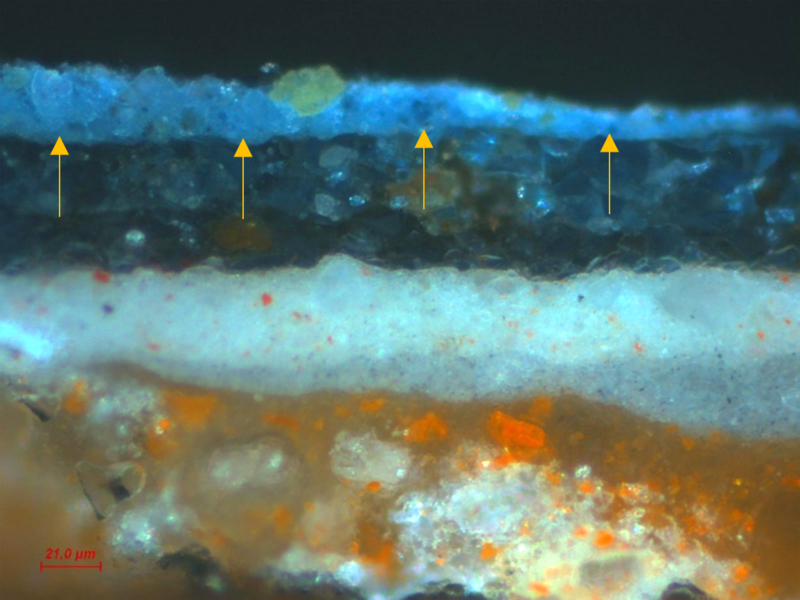
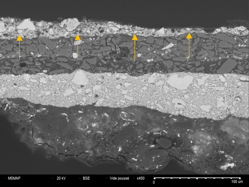
Optical microscopy and SEM-BSE examinations of a paint sample’s stratigraphic section from a painting dating from the early 17th century. On the surface of the original paint layers, a light blue overpaint of Prussian blue and Naples yellow pigments based can be identified.
Organic pigments are studied using Raman spectroscopy.
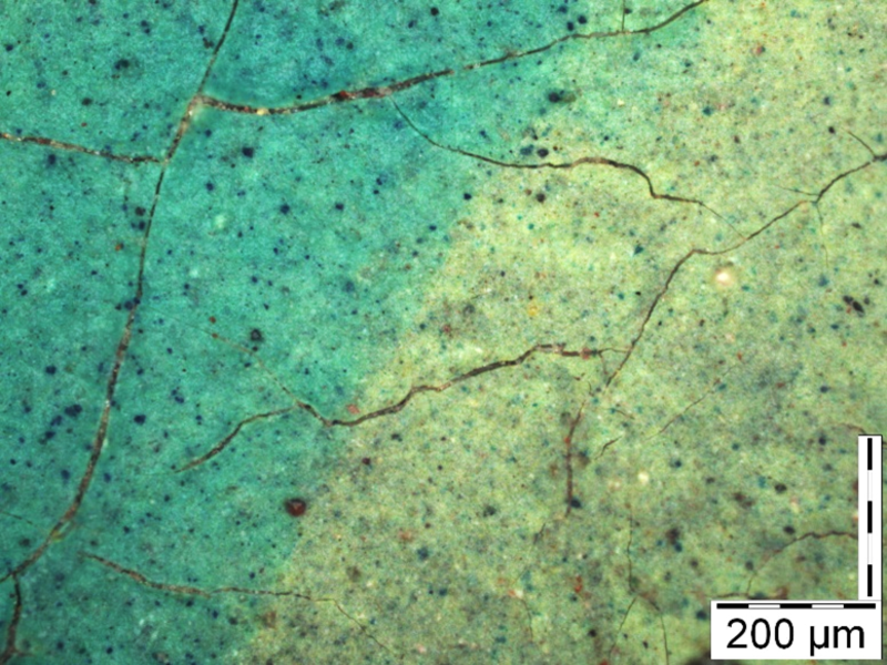
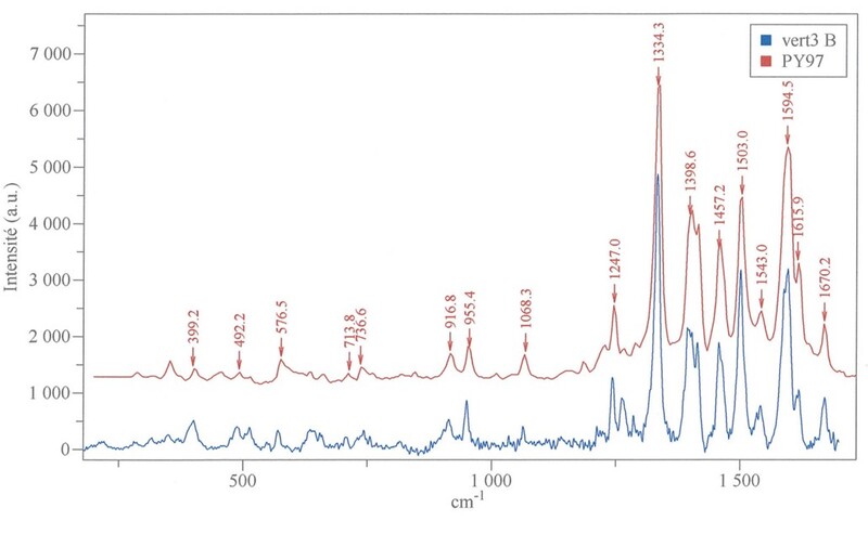
Identification by Raman spectroscopy of a Hansa yellow organic pigment (P.Y. 97) in a painting by Otto Freundlich.
Analysis of organic binders
Analysis of binders, glues, waxes, resins and lacquers are proceeded layer by layer using Fourier transform infrared micro-spectrophotometry.
Study of supports
Studying the support (canvas, paper, cardboard, wooden panels etc.) can also provide essential informations about the period in which the work was produced.
The nature of the fibres making up a canvas is an important chronological indicator.
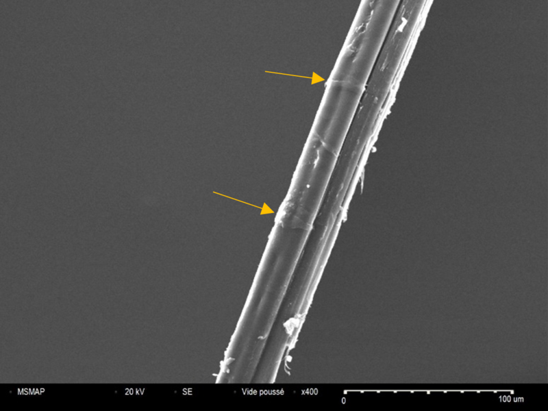
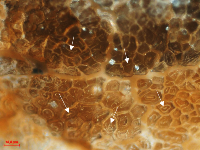
Textile fibres have characteristic morphologies and structures, revealed by optical microscopy and scanning electron microscopy, by means of surface direct observation and on a section.
Flax fibres for example are characterised by well-marked transverse dislocations and nodes, as well as a polygonal fibre shape with sharp points and a narrow lumen.
SCIENTIFIC IMAGING
Revealing the invisible…
Ultraviolet Fluorescence Photography explores the surface of the paint. Overpainting and areas restoration are highlighted. The homogeneity of old or more recent varnishes is also assessed.
Thanks to infrared reflectography, it is possible to identify the underlying layers of a painting and reveal, for example a preparatory drawing, a change in composition or a hidden signature. This examination can prove decisive in the search for the authenticity of a work.
The examination is carried out using very high definition infrared reflectography equipment. The device used produces infrared images of previously unattainable definition with up to several hundred million pixels depending on the format. The quality of the image obtained is more than ten times greater than the definition offered by the equipment available for research into heritage materials. This precision means that the artist’s creative process can be read more clearly since the technology reveals the finest traces of the underlying drawing and the artist’s pentimento.
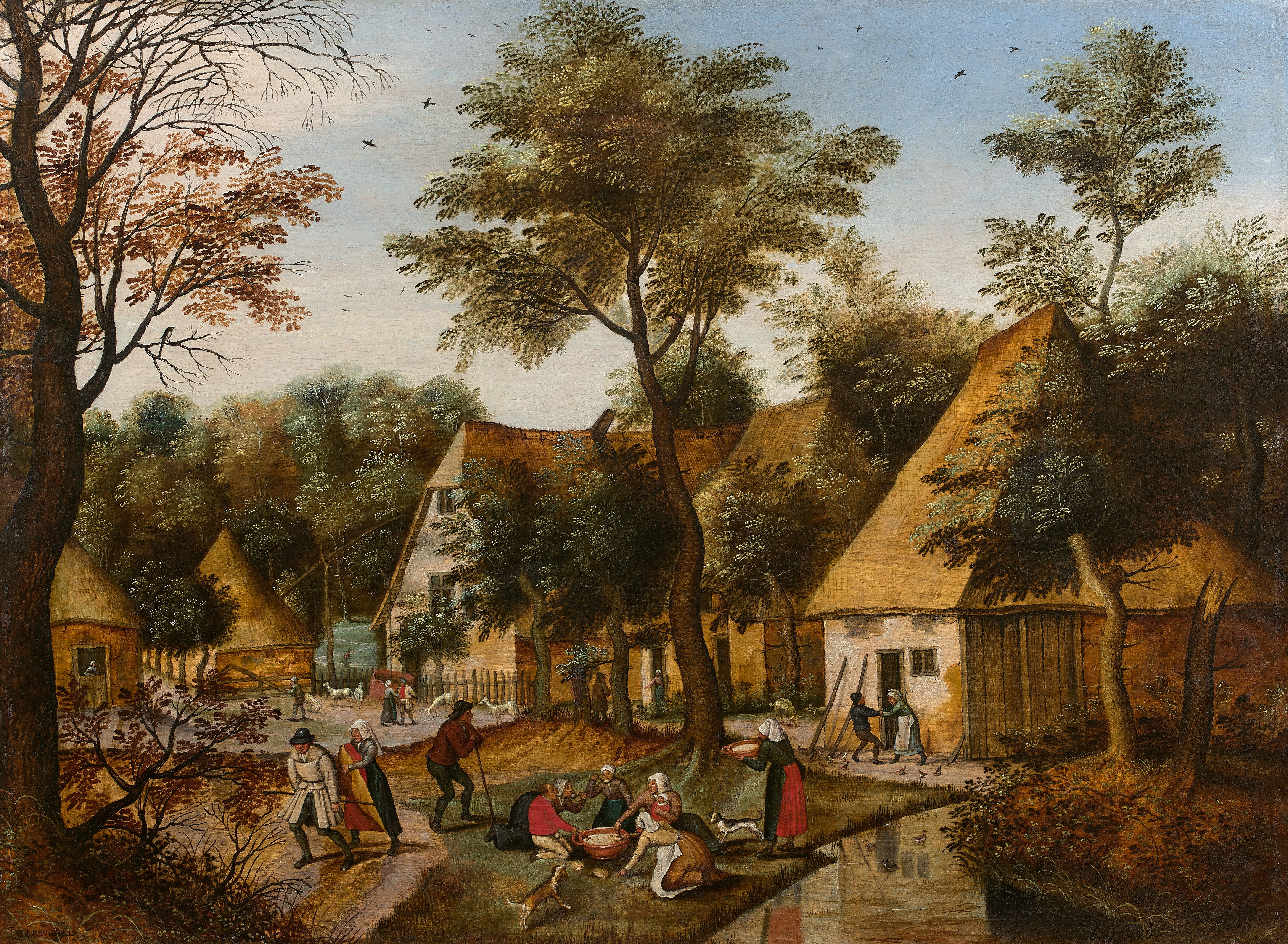
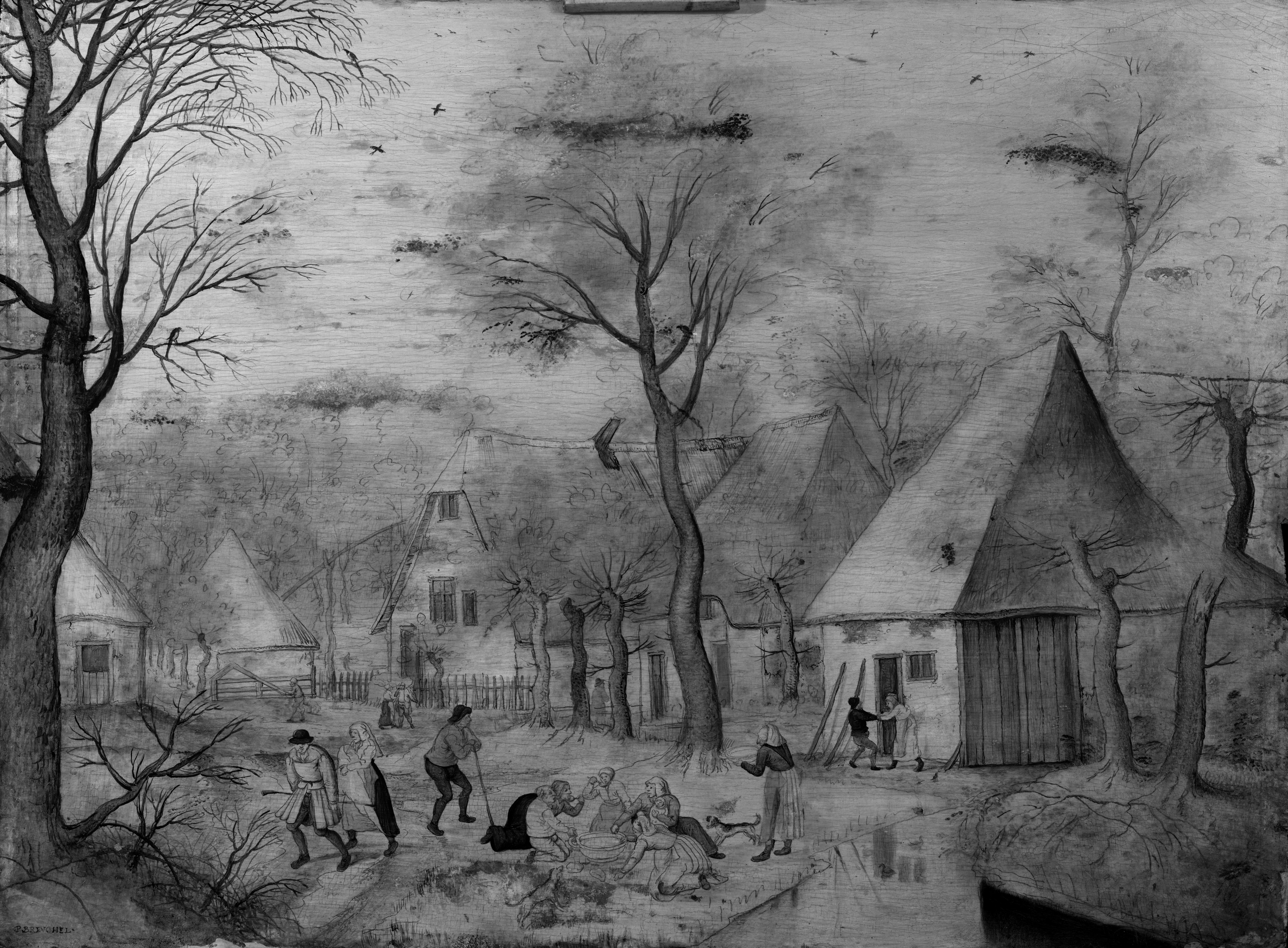




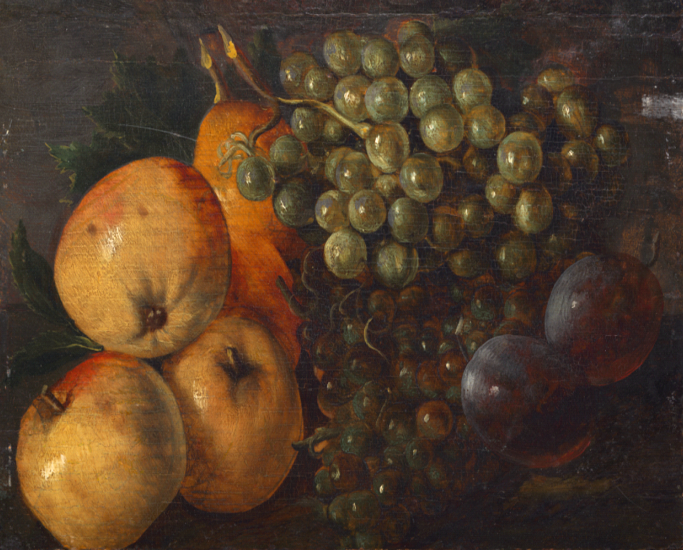
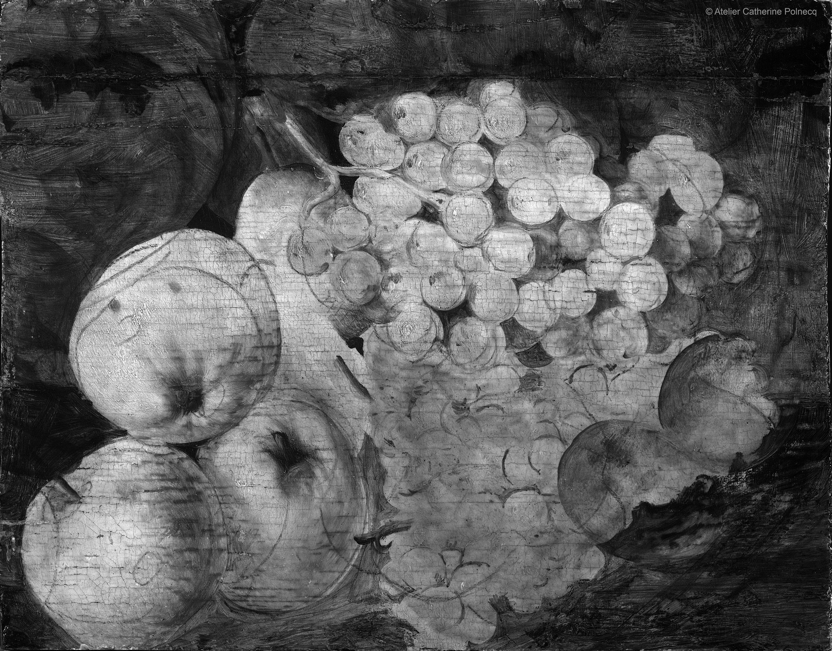
The radiography allows us to explore the deepest layers of a painting. It reveals the general state of conservation of the work, compositional changes, alterations to the support (cracks, tears, etc.) or repairs.
